Lượt truy cập
0486023
Hôm nay:12
Hôm qua:57
Tuần này:140
Tháng này:1749
Tất cả:486023
Đang trực tuyến:3
Hỗ trợ kỹ thuật » Tin chăn nuôi
Chẩn đoán Bệnh dịch tả heo Châu Phi qua bệnh tích (01/09/2018)
African Swine Fever: Pig. There is bloody, mucoid, foamy nasal discharge.           African Swine Fever: Pig, limbs. There is marked hyperemia of the distal limbs.                   African Swine Fever: Pig, perineal skin. There is a large sharply demarcated zone of hyperemia.             African Swine Fever: Pig. There are multiple sharply demarcated foci of cutaneous hemorrhage and/or necrosis; hemorrhagic lesions may contain dark red (necrotic) centers.
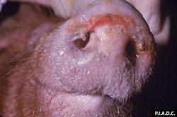
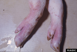
H1. Xoang mũi tiết dịch nhầy, đôi khi có máu.  H 2. Vùng ngoại vi của chân bị sung huyết . H 3. Vùng da mỏng bị sung huyết, xuất huyết . H 4. Vùng trung tâm bị xuất huyết, hoại tử màu đen
 
African Swine Fever: Pig. There are multiple sharply demarcated foci of cutaneous hemorrhage and/or necrosis; hemorrhagic lesions may contain dark red (necrotic) centers.        African Swine Fever: Pig, skin. Necrotic exudate is sloughing from the lesion on the left. There is a rim of hyperemia around the focus of hemorrhage and necrosis (infarct) on the right.               African Swine Fever: Pig, kidney. The cortex contains numerous coalescing petechiae and ecchymoses.             African Swine Fever: Pig, urinary bladder. There are disseminated mucosal petechiae.  
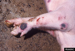
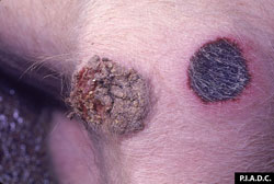
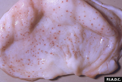
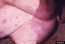
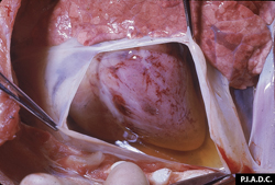
H 5. Vùng chân bị xuất huyết, hoại tử màu đen. H 6. Vùng  bẹn bị xuất huyết, hoại tử màu đen . H 7. Quả thận bị sưng, xuất huyết rất nặng. H 8. Niêm mạc bàng quan xuất huyết lấm chấm hình đinh gim      
 
African Swine Fever: Pig, kidney. Close-up of cortical petechiae.     African Swine Fever: Pig, kidney. Petechiae are disseminated throughout the cortex, and there are larger coalescing pelvic hemorrhages.                African Swine Fever: Pig, heart. There is abundant straw-colored pericardial fluid (hydropericardium), and multifocal epicardial hemorrhage.         African Swine Fever: Pig, lung. The lung is noncollapsed and edematous; there is dorsal hemorrhage and ventral tan consolidation.
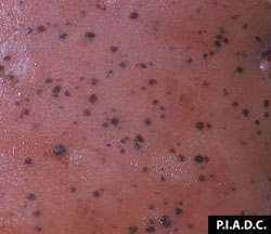
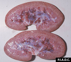
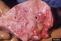
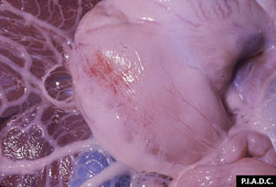
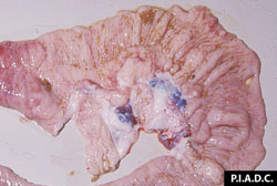
   H 9. Thận xuất huyết điểm đen, hoại tử . H 10.  Vỏ thận xuất huyết điểm hình đinh gim. H 11.  Tim xuất huyết nặng, xoang chứa dịch. H 12.  Phổi viêm, xuất huyết, tích dịch
 
African Swine Fever: Pig, stomach. There is     African Swine Fever: Pig, mandibular lymph node. There is moderate peripheral (medullary) hemorrhage.           African Swine Fever: Pig, stomach. The hepatogastric lymph node is markedly enlarged and hemorrhagic, and the adjacent lesser omentum is edematous.           African Swine Fever: Pig, stomach. The stomach is filled with clotted blood, and the wall is markedly edematous.
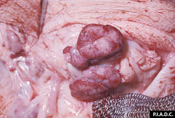
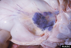
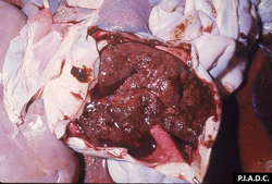
H 13. Dạ dày xuất huyết vệt trên màng niêm mạc. H 14. Hạch lympho xuất huyết đá hoa vân .  H 15. Hạch lympho dạ dày xuất huyết, tụ huyết nặng. H 15.  Xuất huyết nặng màu nâu đen trong dạ dày
 
African Swine Fever: Pig, spiral colon. The colon is distended with bloody contents (due to a hemorrhagic gastric ulcer).     African Swine Fever: Pig, cecum. Mucosa is markedly edematous and hyperemic, and lymph nodes are hemorrhagic.
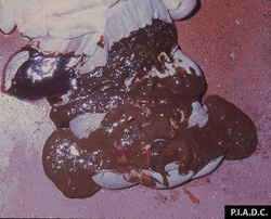
H 16.  Trong ruột chứa máu màu nâu đen.          H 17.  Niêm mạc ruột xuất huyết, hạch ruột xuất huyệt tụ máu       
 
 
ThS . Lê Thanh Tú - Phòng R&D ( sưu tầm)